INTRODUCTION
The NK cells belong to the innate lymphocyte population and are involved in the first line of immune defense system against virally infected and transformed cells [1]. NK cells were initially discerned for their aptitude to kill tumour cells without preparing or previous initiation in divergence to cytotoxic T-cells. Hence, they were named as NK cells. NK cells are extensively dispersed throughout the body in lymphoid and non lymphoid tissues. The highest incidence of NK cells is seen in the lung, then the liver, outlying blood, spleen, bone marrow, lymph nodes, and thymus [2]. Successive discoveries showed the presence of NK cells in other tissues like skin, uterus, salivary gland and adipose tissue [3]. During various physiological and pathological conditions, NK cells secrete an array of cytokines, among which Interferon-γ (IFN-γ) is predominant [4]. Morphologically, they are bone marrow derived large, granular lymphocytes and phenotypically they are Cluster of Differentiation 56+ (CD56+) and CD3- in humans. They are the third largest population of lymphocytes following B and T-cell and represent 10% of total peripheral blood mononuclear cells [5].
Circulating NK cells can be differentiated from liver NK cells by CD16+, presence of chemokine receptors like Chemokine Receptor 1 (CXCR1), CX3CR1 (CD56 dim) and low Natural Killer Cell Receptor (NKG2A), CD27, TNF-Related Apoptosis-Inducing Ligand (TRAIL), perforin, granzyme B, cytokine production and high Antibody Dependent Dell-Mediated Cytotoxicity (ADCC) [2]. In contrast to peripheral NK cells, NK cells inside a vigorous liver display an advanced level of cytotoxicity versus cancer cells and express advanced levels of cytotoxicity mediators [6].The liver also harbours another set of cells in a large percentage called the invariant Natural Killer T (iNKT) cells as compared to any other organ [7].
Glycolipid α-galatosylceramide activates the invariant NKT cells, presented by CD1d which is expressed by various lymphoid and non lymphoid cell types [8]. NK cells in liver are five times higher than in peripheral blood. They play a very vital role in avoidance of Hepatocellular Carcinoma (HCC). They are also considered as potential cell therapy resource for the treatment of HCC [9].
This review highlights the recent advances in understanding the journey and functions of NK cells and their role in various liver diseases.
NK cell development
Human NK cells are present as early as 6 and 15 weeks of gestation in foetal liver and spleen, respectively. Foetal NK cells are functionally immature and they are hyporesponsive compared to adult NK cells. NK cells originate from self-reviving Haematopoietic Stem Cells (HSCs) that reside in the bone marrow. According to recent studies, NK cells can also develop in lymph nodes and liver. NK cell Precursors (NKP) segregates into NK cells however not to other lineages, followed by phenotypic and working NK cell development. Later, NK cells undergo homeostasis. Transcription factors like Erythroblast Transformation Specific-1 (ETS-1), Inhibitor of Deoxyribonucleic Acid (DNA) binding-2 (ID2), Ikaros and PU.1 regulate NK cell development and maturation. GATA-3 and Interferon Regulatory Factor-2 (IRF-2) are involved in maturation of immature NK cells. The cytokine Interleukin-15 (IL-15)-15 has been disclosed to be necessary for NK cell growth, homeostasis and subsistence [5].
Findings by Eissens D et al., have linked the role of 10-colour flow cytometry and recognised seven unique NK cell evolving stages in bone marrow and discovered that NK cell growth is complemented by initial appearance of stimulatory co-receptor CD244. They proposed the seven stages (depicted in [Table/Fig-1]) [10].
- Stage 1: Begins with CD34+, CD117-, CD56-, CD94- cells.
- Stage 2: The ‘Gain’ of CD117 to Stage 1 attains Stage 2 of CD34+, CD117+, CD56-, CD94-.
- Stage 3: A ‘Loss’ of CD34 expression from Stage 2 is Stage 3a (CD34-, CD117+, CD56-, CD94-). Stage Stage 3b (CD34-CD117+CD56+CD94-) is attained with a ‘Gain’ of CD56 from Stage 3a.
- Stage 4: In Stage 4, subsequent to NK cell lineage pledge, cells ‘Gain’ CD94 expression from Stage 3b to grow into undeveloped CD56 bright NK cells or CD34-, CD117+, CD56+, CD94+.
- Stage 5: Stage 5a is attained by a ‘Loss’ of CD117 expression from Stage 4, CD56 dim cells begin to grow, which results into CD34-, CD117-, CD56+, CD94+. Stage 5b (CD34-, CD117-, CD56+, CD94-) results with a ‘Loss’ of CD94 from Stage 5a.

More importantly, both Stage 1 and Stage 2 cells still acquire multi-lineage prospective and consequently comprises of NKPs but they have the ability to provide additional cell lineages. Additional investigation of cord blood, peripheral blood, inguinal lymph node, liver, lymph node, and spleen samples indicated that differential NK cell variation may happen at diverse anatomical sites due to unique appearance profiles of early progress marker CD33 and receptor NKG2A [10].
NK cell functions
Human NK cells in peripheral blood have diverse biological functions which can be classified into two subsets:
- CD56 dim (90%): High cytotoxic activity, and
- CD56 bright (10%): Production of cytokines.
NK cell functioning is controlled by wide range of receptors which are either inhibitory or activating in nature. NK cell receptors identify self Major Histocompatibility Complex (MHC) class 1 molecule and this averts NK cell triggering thereby explaining self-tolerance. The cells which lack self-MHC class 1 molecule activate the NK cells known as ‘missing-self’ hypothesis. Virally infested cells and cancer cells downregulate MHC class 1 expression which helps to avoid killing by cytotoxic T lymphocyte. However, it induces potent stimulatory signals tripping the balance in favour of NK cell activation, which is referred as ‘induced-self recognition’.
NK Cells in Uterus
NK cells are also detected in the uterus known as uterine Natural Killer (uNK) cells. uNK cells lack CD16 but express CD94 and secrete cytokines like Macrophage Inflammatory Protein 1α (MIP1α), Granulocyte Macrophage-Colony Stimulating Factor (GM-CSF), CSF1 and IFN-γ. The uNK cells are also seen in the implantation site and regulate pathogenic T helper 17 (Th17) cells at maternal-foetal border and encourage immune acceptance during pregnancy. Once the target is recognised by NK cells, the two major functions of NK cells are cytotoxicity and cytokine production. NK cells perform ADCC through CD16, as they express a low-affinity Fcγ receptor IIIA. Through apoptosis NK cells kill tumours and virally infected cells. Perforin is a membrane-disrupting protein that is released on NK cell activation and its expression is enhanced by stimulation of IL-2. Granzymes belongs to a line of intrinsically associated serine proteases which are discharged by exocytosis, which along with perforin prompt apoptosis of the target cell. NK cells also express TRAIL and Fas Ligand (FasL) which results in caspase-dependent apoptosis which is associated with death receptors (e.g., FasL/CD95) [5].
NK Cells in Liver Disease
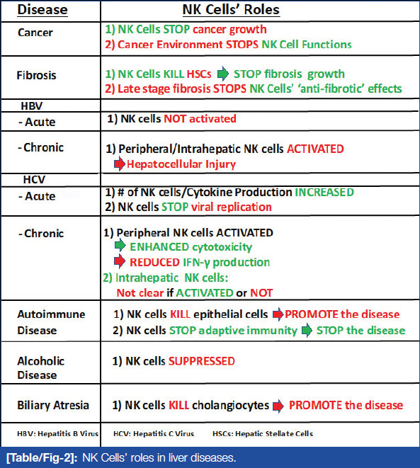
Liver diseases are broadly categorised into viral hepatitis, Alcoholic Liver Disease (ALD), chronic hepatitis like autoimmune liver disease, drug induced liver disease, extrahepatic biliary obstruction, cirrhosis, and Hepatocellular Carcinomas (HCC). [Table/Fig-2] shows these liver disease categories in conjunction with the various roles NK cells play in fighting them.

NK Cells in Viral infection
NK cell has a role as immune-protective agent in viral infection by reducing its number and activity. In various virus infections like Flavi viruses, Human Immunodeficiency Virus (HIV), respiratory syncytial viruses and influenza there is shift from CD56 dim to CD56 bright which causes increased cytokine production [11]. In viral associated asthma, NK cells contribute to Immunoglobulin E (IgE) mediated immune response and cause resolution of acute allergic airway inflammation. They migrate from circulation towards tissues like lung and lymphoid organs due to antigenic stimulation [12].
NK cells in viral hepatitis: The scientists have suggested that NK cells play a critical role in defense against viral hepatitis. Globally, viral hepatitis has been the leading source of end-stage liver disease and hepatocellular carcinoma. Since the mid-1960s, five hepatitis viruses, types A, B, C, D and E, have been identified. More than half a billion people are persistent carriers of the Hepatitis B Virus (HBV), the Hepatitis C Virus (HCV), or the Hepatitis Delta Virus (HDV).
NK cells are known to play an important role in viral infection. Initial findings claimed that HCV obstructs NK cell tasks and getaways from immune reconnaissance of NK cells resulting in lingering infection. During acute infection, NK cells are activated by INF-α/β, IL-12, IL-15, IL-18 which inturn cause killing of HCV infected hepatocytes [13]. It has been observed that peripheral NK cells are also triggered during continuing HCV infection which shows an upsurge cytotoxicity with raised expression of NKG2D, NK p46, TRAIL and STAT1 which contributes to liver injury. Decrease IFN-γ production may facilitate inability to clear HCV resulting in chronic stage [14].
Hepatitis B virus (HBV): Acute HBV infection in early stage shows upregulation of IL-10 in absence of IFN-α/β and IL-15 induction which contributes to inhibition of peripheral NK cell functions. Chronic HBV infection causes liver inflammation by activation of peripheral and intrahepatic NK cells induced by IFN-α, IL-12, 15, 18. Activated NK cells cause hepatocellular damage. However its role in control of HBV replication is questionable [15,16].
Ghosh S et al., study showed that NK cells cause hepatic harm and aid in viral resolve during advancement of hepatitis B. This is mediated by insufficient IFN-γ production and NK mediated, perforin dependent depletion of CD4+T cells which causes liver damage and HBV persistence contributing to advance liver damage. This finding demonstrated that immunotherapy that combines INF-α and therapeutic weakening of NK cells or obstruction of NK triggering receptors can be effective in HBV suppression [17].
NK Cells in COVID-19
NK cells play an important role in moderating the immune response in Coronavirus Disease-2019 (COVID-19) patients. There is decrease in NK cell number and its function, resulting in reduced clearance of activated and infected cells, and unhindered increase of tissue-damaging irritation markers. COVID-19 infection tilts the immune reaction towards a tremendously inflammatory phenotype. Re-establishment of NK cell effector functions has the prospective to appropriate the delicate immune balance necessary to successfully overcome COVID-19 infection [12].
NK Cell in Autoimmune Disease
In autoimmune diseases like immune encephalomyelitis and multiple sclerosis, there is a reduction of number of NK cell with decrease cytotoxic function. It is also observed that cytotoxicity of NK cells can augment an autoimmune disease by destruction of cells in a target organ [18].
In type 1 diabetes, NK p46, the activating NK cell receptor binds to a known ligand on β-cell of pancreas and effectively killing them [19].This is essential for development of type I diabetes. Various other studies revealed low expression of NK p30, NK p46 receptors in patients with long standing type I diabetes. They also displayed reduced perforin massenger Ribonucliec Acid (mRNA) expression along with decreased lysis activity by NK cells [20]. This was thought to be a consequence of diabetes rather than a cause.
In other autoimmune diseases role of NK cells is either disease promoting or disease controlling. Like in rheumatoid arthritis, there is NK cell accumulation in synovial fluid by secretion of IFN-γ via CD56 bright [21].
In systemic lupus erythematous, function of NK cells is reduced and there is shift from CD56 dim to CD56 bright subset [22]. Abnormalities in number and function in this inflammatory condition play protective or disease controlling role. NK cells also have a crosstalk with other immune cells like macrophages and Treg cells [23]. Treg cells have been seen to subdue NK cells via IL-21 arbitration in autoimmune illnesses. Exact mechanism and role of NK cell in autoimmune disease as a friend or foe is debatable. This also suggests the possibility of role of NK cell in causing remission and exacerbation of these autoimmune conditions [24].
Role of NK cells in Liver
NK cells in liver were first studied in mouse. Liver resident NK cells were DX5+ and DX5-. Hepatic NK cells were approximately half enriched in DX5- NK cells. CD49a is considered to be specific marker for liver DX5- NK cells, rarely expressed by liver DX5+ NK cells [25]. The CD49a+DX5- NK cells were present in liver sinusoid blood, neither in the afferent nor efferent blood vessels of the liver [26]. But CD49a-DX5+ is present in all blood sources. In human liver CD56 bright NK cells expressed CD49a and these CD49a+ NK cells express transcription factors like -bet+ Eomes- (T-box expressed in T-cells and Eomesodermin) [27]. In human liver-resident NK cells surface markers are CD56 bright CD16-, CXCR6+, CCR5+, CD69+, CD49a+/-, CX3CR1-, CD57-, CD49e-. Effector molecules include TRAIL, granzyme K, IFN-γ, TNFα, GM-CSF. Human liver NK cell lacks T-bet expression and it highly expresses Enomes [28].
NK cell is a key component of the innate immune system in the liver. They act by fabrication of cytokines, killing cancer cells, pathogens, strained hepatocytes and HSCs. NK cells acts as a governing cell which influences other cells in the vicinity like Dendritic Cells (DCs), T-cells, B-cells, Kupffer cells and endothelial cells by producing various cytokines like INF-γ, TNF-α, IL-10 and TLR ligands, chemokines and growth factors. T regulatory cells and activated HSCs can inhibit NK cell formation via inhibitory cytokines predominantly TGF-β, IL-10. Triggered NK cells mark HSCs, hepatocytes, and cholangiocytes and achieve range of vital functions in the pathogenesis of liver illness [13].
NK Cell in Alcoholic Liver Disease (ALD)
Alcohol consumption contributes to an increase in NK cell cytotoxicity in normal individuals which may contribute to development of ALD. However, it is observed that patients with ALD show decrease NK cell numbers and reduce cytotoxic activity [29]. Chronic exposure of alcohol causes reduced expression of NKG2D, TRAIL and IFN-γ on NK cells. This results in decreased antiviral, antifibrotic and antitumour effects of NK cells which contributes to susceptibility to infection, accelerated liver fibrosis and HCC in patients with liver alcoholic disease.
Obesity adds to decrease circulating NK cells with lower level of cytotoxicity [30]. However, Kahraman A et al., study reported NK cell associated cytotoxic mediators (such as TRAIL, NKG2D and MIA/B mRNAs), and number of hepatic NK cells were strikingly amplified in overweight patients with Non Alcoholic Steatohepatitis (NASH)and to smaller degree with non alcoholic fatty liver when rivalled to normal individuals [31]. In NASH patients, the expression of MICA/B mRNAs correlates with the non alcoholic fatty liver disease (NAFLD) activity score and hepatocyte apoptosis thus, NK cells are triggered due to the raised levels of numerous cytokines (e.g., IL-12, IL-18, and IFN-γ) and ligands (e.g., MICA/B) and contribute to the pathogenesis of NASH [31,32].
Fibrosis and Cirrhosis
NK cells plays important role in controlling liver fibrosis which were recently seen in HCV patients by several clinical studies [33]. The first study demonstrated, human NK cells contributed to production of TRAIL and FasL which resulted in killing of the activated primary human HSCs in vitro. Second, activation of NK cell mediated killing of human HSCs through NKG2D and NK p46 activating receptors. Third, contribution of INF-α in patient with HCV increased the ability of NK cells to kill primary human HSCs. Fourth, cytotoxicity of NK cells were isolated from HCV patients against primary human HSCs demonstrated inverse correlation with stage of liver fibrosis. Fifth, lymphocytes of HCV patients were transfected with the help of KIR small interfering RNAs (siRNAs) inhibited the activation of human HSCs [34]. Lastly, buildup of NK p46 high NK cells in liver was contrariwise linked with the fibrosis phase of HCV patients. All these discoveries propose that NK cells perform a vital role in reducing liver fibrogenesis. Though, chronic alcohol consumption can reduce the antifibrotic function of NK cells and raised levels of TGF-β are associated with final stage liver fibrosis, which leads to advancement of liver fibrogenesis [35].
Autoimmune Liver Illness
Various human autoimmune diseases of liver like autoimmune hepatitis, Primary Biliary Cirrhosis (PBC), primary sclerosing cholangitis reduces the NK cell functions. In the pathogenesis of these disorders NK cells play dual roles [36]. NK cells cause destruction of biliary epithelial cells via a TRAIL-dependent mechanism and by cytokine production which promotes adaptive immune response and progression of disease. In contrast, NK cells may also cause regression of PBC by production of IL-10 and killing of T-cells and DCs [37].
NK Cells in Biliary Disease
Biliary atresia, an advanced fibro-obliterative cholangiopathy with unidentified aetiology, disturbs the biliary tree in infants and upsets bile flow from liver to the intestine. Investigational simulations implied that NK cells destroy cholangiocytes via NKG2D [38]. Postnatal absence of T regulatory cells in these patient allow hepatic DCs to act unopposed in NK cell activation and cholangiocyte destruction [35,39].
Liver Cancer
NK cells are enriched in healthy liver and they play an important role in the immune surveillance for tumours which is mediated via the production of perforin, granzyme, TRAIL, IFN-γ [40]. In HCC patients, the number of peripheral CD56dim CD16+ NK cells decreases with impaired cytotoxic activity and IFN-γ production [41].Several mechanisms involved in pathogenesis of HCC are NK cell malfunction, decrease in its number resulting in escape of tumour cells from immune surveillance.
Fibroblasts present within HCC produces Indoleamine 2,3-dioxygenase (IDO) and Prostaglandin E2 (PGE2) which trigger NK cells dysfunction and downregulates activating NK receptors [42]. In HCC patients, Myeloid-Derived Suppressor Cells (MDSCs) interact with NK cells and contribute to decreased cytotoxicity and cytokine production of NK cells [43].
NK cells as therapeutic targets for the treatment of liver disease
In view of the important role of NK cell in pathogenesis of liver disorders, there is strong possibility of using hepatic NK cells as potential therapeutic target. Several approaches include cytokine treatment to increase the cytotoxicity of NK cell, antibodies to modulate NK cytotoxic function and use of agonist of NK cell activating receptors and adoptive transfer of NK cells. IL-12 and IL-18 have been displayed to successfully obstruct liver carcinogenesis by enhancing NK cell anticancer task. Recombinant IL-2 and IL-15 triggers both NK cell and CD8+ T-cell without exciting Tregs [44].These are presently being verified for haematological malignancies. MiR-182 has been shown to rise NK cell cytotoxicity by controlling expression of NKG2D and NKG2A in HCC [45].
In this new era of immunotherapy for cancer, several inhibitory check points are targeted on NK cell through blocking monoclonal antibodies (mAbs) [46]. Killer cell immunoglobin like receptors KIR are expressed on NK cells and is also present on T-cell in minor amounts. Antibodies against anti-KIR or inhibitory KIR showed promising effect in haematological cancer. NKG2A is currently been tested in solid tumours [47]. NK cells also express PD1. mAbs blocking PD-1/PD-L1 can be the future therapeutic target for liver cancer [48]. More recently genetic engineering with different technological approach like bi- and tri-specific killer engagers (BiKEs and TriKEs) or Chimeric Antigen Receptors (CARs) showed improved ability of NK cells to infiltrate tumour tissue [49]. This can be used as NK cell transfer therapies in liver cancer in future.
NK cells role in liver transplant
Liver transplantation or LT can save lives for patients with acute and persistent liver malfunction. It is the last treatment option for patients with:
a) Major complications caused by final stage chronic liver disease;
b) Sudden failure of a previously healthy liver.
Transplant receivers need lifelong immuno-suppression to avert immune responsiveness directed versus the donor organ, causing in graft refusal. NK cells role in organ transplantation has been inadequately defined due to contradictory clinical and experimental statistics. However, the liver contains main resident populations of immune cells, particularly enriched with NK cells, γ-δ T-cells, and NKT cells. Transplantation of liver thus ends in a rare assembly of recipient and donor immune systems. This potentially inflammatory meeting results in attenuated immune response leading to less requirement of immunosuppression. Thus, liver NK cell play an important role in tolerance induction and post liver transplant outcome [50].
CONCLUSION(S)
Recent studies have facilitated us to increase an understanding of NK cell in relations of its development, role, distinct phenotypical features and receptor interactions. Their assignment in confronting cancer cells is well recognised. The identification of liver resident NK cells have opened various dimensions in understanding the pathogenesis of liver diseases like hepatitis, ALD, autoimmune liver disease, non alcoholic steatohepatits, biliary atresia and HCC. Interestingly, they play an important role in induction of tolerance post liver transplant. Over the last two decades, research in NK cells has gained some traction in therapeutics for the diseases of liver and other organs. However, how to deploy NK cells for the therapy of diseases remains in infancy as most of this work is still in the preclinical stage. A nationwide or global NK strategy is therefore a must to improve ailment therapies further.
REFERENCES
[1] Vivier E, Tomasello E, Bartin M, Walzer T, Ugoliniet S. Functions of natural killer cells. Nat Immunol. 2008;9:503-10.
[2] Zhigang T, Yongyan C, Bin G. Natural killer cells in liver disease. Hepatology. 2013;(574):1654-62.
[3] O’Sullivan T, Rapp M, Fan X, Weizman O, Bhardwaj P, Adams N, et al. Adipose-resident group 1 innate lymphoid cells promote obesity-associated insulin resistance. Immunity. 2016;45:428-41.
[4] Vivier E, Raulet D, Moretta A, Caligiuri A, Zitvogel L, Lanier L, et al. Innate or adaptive immunity? The example of natural killer cells. Science. 2011;331:44-49.
[5] Mandal A, Viswanathan C. Natural killer cells: In health and disease. HematolOncol Stem Cell Ther. 2015;8(2):47-55.
[6] Ishiyama K, Ohdan H, Ohira M, Mitsuta H, Arihiro K, Asahara T. Difference in cytotoxicity against hepatocellular carcinoma between liver and periphery natural killer cells in humans. Hepatology. 2006;43:362-72.
[7] Haider R, Seki S, Weerasinghe A, Kawamura T, Watanabe H, Abo T. Characterization of NK cells and extrathymic T cells generated in the liver of irradiated mice with a liver shield. Clin Exp Immunol. 1998;114:434-47.
[8] Sullivan B, Kronenberg M. Activation or anergy: NKT cells are stunned by alpha-galactosylceramide. J Clin Invest. 2005;115:2328-29.
[9] Sun H, Sun C, Tian Z, Xiao W. NK cells in immunotolerant organs. Nature, Cellular & Molecular Immunology. 2013;10(3):202-12.
[10] Eissens D, Spanholtz J, van der Meer A, van Cranenbroek B, Dolstra H, Kwekkeboom J, et al. Defining early human NK cell developmental stages in primary and secondary lymphoid tissues. PLoS One. 2012;7(2):e30930.
[11] Lunemann S, Malone D, Hengst J, Port K, Grabowski J, Deterding K, et al. Compromised function of natural killer cells in acute and chronic viral hepatitis. J Infect Dis. 2014;209(9):1362-73.
[12] van Eeden C, Khan L, Osman M, Cohen T. Natural killer cell dysfunction and its role in COVID-19. Int J Mol Sci. 2020;21(17):6351.
[13] Tian Z, Chen Y, Gao B. Natural killer cell in liver disease. Hepatology. 2013;57(4):1654-62.
[14] Ahlenstiel G, Titerence R, Koh C, Edlich B, Feld J, Rotman Y, et al. Natural killer cells are polarized toward cytotoxicity in chronic hepatitis C in an interferon-alfa-dependent manner. Gastroenterology. 2010;138:325-35.e3-2.
[15] Dunn C, Brunetto M, Reynolds G, Christophides T, Kennedy P, Lampertico P, et al. Cytokines induced during chronic hepatitis B virus infection promote a pathway for NK cell-mediated liver damage. J Exp Med. 2007;204:667-80.
[16] Zhang Z, Zhang S, Zou Z, Shi J, Zhao J, Fan R, et al. Hypercytolytic activity of hepatic natural killer cells correlates with liver injury in chronic hepatitis B patients. Hepatology. 2011;53:73-85.
[17] Ghosh S, Nandi M, Pal S, Chakraborty B, Khatun M, Bhowmick D, et al. Natural killer cells contribute to hepatic injury and help in viral persistence during progression of hepatitis B e-antigen-negative chronic hepatitis B virus infection. Clinical Microbiology and Infection. 2016;733.e9-19.
[18] Hao J, Liu R, Piao W, Zhou Q, Vollmer T, Campagnolo D, et al. Central nervous system (CNS)-resident natural killer cells suppress Th17 responses and CNS autoimmune pathology. J Exp Med. 2010;207(9):1907-21.
[19] Gur C, Enk J, Kassem S, Suissa Y, Suissa Y, Magenheim J, et al. Recognition and killing of human and murine pancreatic beta cells by the NK receptor NKp46. J Immunol. 2011;187(6):3096-103.
[20] Rodacki M, Svoren B, Butty V, Besse W, Laffel L, Benoist C, et al. Altered natural killer cells in type 1 diabetic patients. Diabetes. 2007;56(1):17-85.
[21] Pazmany L. Do NK cells regulate human autoimmunity? Cytokine. 2005;32(2):76-80.
[22] Hervier B, Beziat V, Haroche J, Mathian A, Lebon P, Ghillani-Dalbin P, et al. Phenotype and function of natural killer cells in systemic lupus erythematosus: Excess interferon-c production in patients with active disease. Arthritis Rheum. 2011;63(6):1698-706.
[23] Nedvetzki S, Sowinski S, Eagle R, Harris J, Vély F, Pende D, et al. Reciprocal regulation of human natural killer cells and macrophages associated with distinct immune synapses. Blood. 2007;109(9):3776-85.
[24] Zhou Z, Zhang C, Zhang J, Harris J, Vély F, Pende D, et al. Macrophages help NK cells to attack tumour cells by stimulatory NKG2D ligand but protect themselves from NK killing by inhibitory ligand Qa-1. PLoS One. 2012;7(5):e36928.
[25] Peng H, Jiang X, Chen Y, Sojka D, Wei H, Gao X, et al. Tissue-resident natural killer (NK) cells are cell lineages distinct from thymic and conventional splenic NK cells. Elife. 2014;3:e01659.
[26] Yokoyama W, Sojka D, Peng H, Tian Z.Tissue-resident natural killer cells. Cold Spring HarbSymp Quant Biol. 2013;78:149-56.
[27] Marquardt N, Beziat V, Nystrom S, Hengst J, Ivarsson M, Kekäläinen E, et al. Cutting edge: Identification and characterization of human intrahepatic CD49a+ NK cells. J Immunol. 2015;194:2467-71.
[28] Aw Yeang H, Piersma S, Lin Y, Yang L, Malkova O, Miner C, et al. Cutting edge: Human CD49e- NK cells are tissue resident in the liver. J Immunol. 2017;198:1417-22.
[29] Laso F, Almeida J, Torres E, Vaquero J, Marcos M, Orfao A. Chronic alcohol consumption is associated with an increased cytotoxic profile of circulating lymphocytes that may be related with the development of liver injury. Alcohol Clin Exp Res. 2010;34:876-85.
[30] O’Shea D, Cawood T, O’Farrelly C, Lynch L. Natural killer cells in obesity: Impaired function and increased susceptibility to the effects of cigarette smoke. PLoS One. 2010;5:e8660.
[31] Kahraman A, Schlattjan M, Kocabayoglu P, Yildiz-Meziletoglu S, Schlensak M, Fingas C, et al. Major histocompatibility complex class I-related chains A and B (MIC A/B): A novel role in nonalcoholic steatohepatitis. Hepatology. 2010;51:92-102.
[32] Bertola A, Bonnafous S, Anty R, Patouraux S, Saint-Paul M, Iannelli A, et al. Hepatic expression patterns of inflammatory and immune response genes associated with obesity and NASH in morbidly obese patients. PLoS One. 2010;5:e13577.
[33] Gur C, Doron S, Kfir-Erenfeld S, Abu-Tair L, Safadi R, Mandelboim O. NKp46-mediated killing of human and mouse hepatic stellate cells attenuates liver fibrosis. Gut. 2012;61:885-93.
[34] Muhanna N, Abu Tair L, Doron S, Amer J, Azzeh M, Mahamid M, et al. Amelioration of hepatic fibrosis by NK cell activation. Gut. 2011;60:90-98.
[35] Jeong W, Park O, Suh Y, Byun J, Park S, Choi E, et al. Suppression of innate immunity (natural killer cell/interferon-gamma) in the advanced stages of liver fibrosis in mice. Hepatology. 2011;53:1342-51.
[36] Tian Z, Gershwin M, Zhang C. Regulatory NK cells in autoimmune disease. J Autoimmun. 2012;39:206-15.
[37] Schleinitz N, Vely F, Harle J, Jean E, Harlé J, Rossi P, et al. Natural killer cells in human autoimmune diseases. Immunology. 2010;131:451-58.
[38] Shivakumar P, Sabla G, Whitington P, Chougnet C, Bezerra J. Neonatal NK cells target the mouse duct epithelium via Nkg2d and drive tissue-specific injury in experimental biliary atresia. J Clin Invest. 2009;119:2281-90.
[39] Saxena V, Shivakumar P, Sabla G, Mourya R, Chougnet C, Bezerra J. Dendritic cells regulate natural killer cell activation and epithelial injury in experimental biliary atresia. Sci Transl Med. 2011;3:102ra94.
[40] Subleski J, Wiltrout R, Weiss J. Application of tissue-specific NK and NKT cell activity for tumour immunotherapy. J Autoimmun. 2009;33:275-81.
[41] Cai L, Zhang Z, Zhou L, Wang H, Fu J, Zhang S, et al. Functional impairment in circulating and intrahepatic NK cells and relative mechanism in hepatocellular carcinoma patients. Clin Immunol. 2008;129:428-37.
[42] Li T, Yang Y, Hua X, Wang G, Liu W, Jia C, et al. Hepatocellular carcinoma associated fibroblasts trigger NK cell dysfunction via PGE2 and IDO. Cancer Letters. 2012;318(2):154-61.
[43] Hoechst B, Voigtlaender T, Ormandy L, Gamrekelashvili J, Zhao F, Wedemeyer H, et al. Myeloid derived suppressor cells inhibit natural killer cells in patients with hepatocellular carcinoma via the NKp30 receptor. Hepatology. 2009;50(3):799-807.
[44] Thaysen-Andersen M, Chertova E, Bergamaschi C, Moh E, Chertov O, Roser J, et al. Recombinant human heterodimeric IL-15 complex displays extensive and reproducible N- and O-linked glycosylation. Glycoconj J. 2016;33:417-33.
[45] Abdelrahman M, Fawzy I, Bassiouni A, Gomaa A, Esmat G, Waked I, et al. Enhancing NK cell cytotoxicity by miR-182 in hepatocellular carcinoma. Hum Immunol. 2016;77:667-73.
[46] Marcenaro E, Notarangelo L, Orange J, Vivier E. Editorial: NK cell subsets in health and disease: New developments. Front Immunol. 2017;8:1363.
[47] Zaghi E, Calvi M, Marcenaro E, Mavilio D, Di Vito C. Targeting NKG2A to elucidate natural killer cell ontogenesis and to develop novel immune-therapeutic strategies in cancer therapy. J Leukoc Biol. 2019;6:1243-51.
[48] Della Chiesa M, Pesce S, Muccio L, Quatrini L, Munari E, Vacca P, et al. Features of memory-like and PD-1(+) human NK cell subsets. Front Immunol. 2016;7:351.
[49] Daher M, Rezvani K. Next generation natural killer cells for cancer immunotherapy: the promise of genetic engineering. Curr Opin Immunol. 2018;51:146-53.
[50] Harmon C, Sanchez-Fueyo A, Farrelly O, Houlihan D. Natural killer cells and liver transplantation: Orchestrators of rejection or tolerance? American Journal of Transplantation. 2016;16:751-57.


